Surface and Cross Section Anatomy of the Kidney
Image Description:
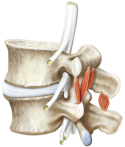
Surface and Cross Section Anatomy of the Kidney.
Image File Sizes:
|
Size |
Pixels |
Inches (@300dpi) |
cm (@300dpi) |
|
Small |
600 x 430px |
2.0 x 1.4” |
5.1 x 3.6cm |
|
Medium |
1200 x 860px |
4.0 x 2.9” |
10.2 x 7.3cm |
|
Large |
2400 x 1721px |
8.0 x 5.7” |
20.3 x 14.6cm |
|
X-Large |
4000 x 2868px |
13.3 x 9.6” |
33.9 x 24.3cm |
|
Maximum |
7554 x 5416px |
25.2 x 18.1” |
64 x 45.9cm |
Anatomy Visible in the Medical Illustration Includes:
Kidney, cortex, fibrous capsule, major calyxes, renal vein, renal artery, ureter, renal papilla, minor calyxes, medulla or renal pyramids, arcuate vein, arcuate artery, interlobular artery, interlobular vein.
Image created by:
We Also Recommend