Thoracic Vertebra in Superior View
Image Description:
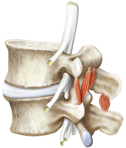
Illustration of a thoracic vertebra in superior view. The thoracic spine refers to the middle portion of the spine that connects with the cervical spine in the neck and the lumbar spine. Made up of twelve vertebrae they are labelled T1 to T12. Between each of the vertebrae are intervertebral discs. The first two thoracic vertebra have been cross sectioned to show the inside of the bone.
Image File Sizes:
|
Size |
Pixels |
Inches (@300dpi) |
cm (@300dpi) |
|
Small |
591 x 600px |
2.0 x 2.0” |
5.0 x 5.1cm |
|
Medium |
1182 x 1200px |
3.9 x 4.0” |
10.0 x 10.2cm |
|
Large |
2375 x 2412px |
7.9 x 8.0” |
20.1 x 20.4cm |
Anatomy Visible in the Medical Illustration Includes:
Thoracic vertebra, costal facet, spinous process, lamina, pedicle, inferior costal facet, superior costal facet, transverse process, superior articular facet, superior vertebral notch, vertebral body, vertebral foramen
Image created by:
We Also Recommend