Upper and Lower Airway Anatomy (Child)
Image Description:
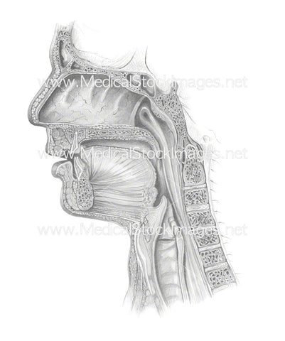
Illustration showing the upper and lower airway of a child. The respiratory tract is divided into upper and lower airways, separated by the larynx/vocal cords. The upper airway (nose, sinuses, pharynx, larynx) warms, filters, and humidifies air. The lower airway (trachea, bronchi, bronchioles, lungs) conducts air to the alveoli for gas exchange. The diaphragm is a large, dome-shaped muscle located below the lungs that serves as the primary muscle for respiration. It contracts and flattens to create a vacuum that pulls air into the lungs, and relaxes to force air out.
Image File Sizes:
|
Size |
Pixels |
Inches |
cm |
|
Small |
600x950px |
2x3” @300dpi |
5x8cm @300dpi |
|
Medium |
1200x1900px |
4.0x6.0” @300dpi |
10x16cm @300dpi |
|
Large |
2400x3800px |
8.0x12” @300dpi |
20.3x32.17cm @300dpi |
|
X-Large |
3600x5700px |
12x12” @300dpi |
30.48x48.26cm @300dpi |
Anatomy Visible in the Medical Illustration Includes:
Child, boy, mouth, nose, nasal cavity, pharynx, larynx, trachea, lungs, tongue, epiglottis, mandible, vocal cords, esophagus, oesophagus, upper airway, lower airway, upper respiratory tract, diaphragm.
Image created by:
We Also Recommend