Bone Healing Stages
Image Description:
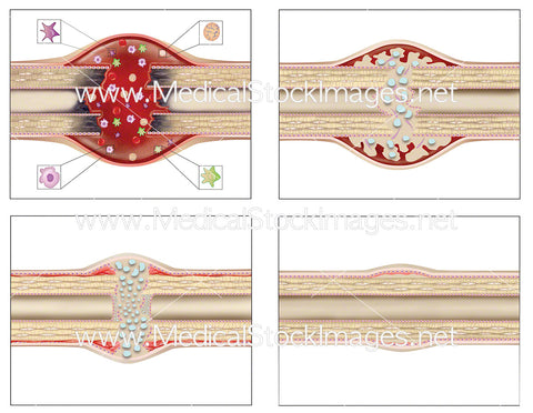
Illustration showing bone healing stages that occur after a fracture or break. The healing stages include an inflammatory stage, repair stage (soft callus formation and hard callus formation) and late remodelling stage. In the inflammatory stage, a hematoma develops during the first hours and days. Four types of inflammatory cells, macrophages, fribroblasts, mesenchymal stem cells and polymorphonuclear cells, enter the bone and form granulation tissue which gradually replaces the hematoma. Osteoclasts remove necrotic one at the ends of the break or fracture. Soft callus is formed around the break after about 2-3 weeks after the injury. Over the next 3-4 months, soft callus is replaced by hard callus which unites the two ends of the break, joining them with new bone. Finally, when the fracture is woven together with new bone, the bone is replaced by lamellar bone until it has returned completely to its original composition. This process can take from 3 months to several years, depending on factors such as the severity and size of the fracture.
Image File Sizes:
|
Size |
Pixels |
Inches (@300dpi) |
cm (@300dpi) |
|
Small |
600 x 465px |
2.0 x 1.6” |
5.1 x 3.9cm |
|
Medium |
1200 x 930px |
4.0 x 3.1” |
10.2 x 7.9cm |
|
Large |
2400 x 1860px |
8.0 x 6.2” |
20.3 x 15.7cm |
|
X-Large |
3757 x 2911px |
12.5 x 9.7” |
31.8 x 24.7cm |
Anatomy Visible in the Medical Illustration Includes:
Bone, break, fracture, healing, periosteum, necrosis, endosteum, fibroblasts, polymorphonuclear cells, macrophages, mesenchymal stem cells, hematoma, cartilage, osteoblasts, soft callus, hard callus, woven bone, lamellar bone
Image created by:
We Also Recommend

