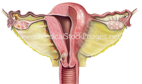
Uterus Cross-Section with Tumour
Image Description:
The body of the uterus is shown with half as surface anatomy and half as a cross section. The image includes the vagina and the fallopian tubes and ovaries. A tumour is shown in the isthmus of uterus area.
Image File Sizes:
|
Size |
Pixels |
Inches (@300dpi) |
cm (@300dpi) |
|
Small |
600 x 358px |
2.0 x 1.2" |
5.1 x 3.0cm |
|
Medium |
1200 x 715px |
4.0 x 2.4" |
10.2 x 6.1cm |
|
Large |
2400 x 1430px |
8.0 x 4.8" |
20.3 x 12.1cm |
|
X-Large |
2933 x 1748px |
9.8 x 5.8" |
24.8 x 14.8cm |
Anatomy Visible in the Medical Illustration Includes:
Body of uterus, broad ligament, ovarian ligament, ovary, round ligament, mesovarium, uterine tube, fundus of uterus, perimetrium, isthmus, ampulla, suspensory ligament, cervix, cervical canal, fimbriae, infundibulum, vagina, vaginal rugae, cervix, endometrium, fallopian tubes, ovaries.
Image created by:
We Also Recommend

